What Is Nipah Virus?
Nipah Virus has gained everyone's attention when the world has just forgotten about the deadly COVID 19. It’s high time for everyone in the world to know about Nipah, at least its mode of transmission and preliminary signs- as this knowledge can be totally lifesaving!
2/4/20264 min read
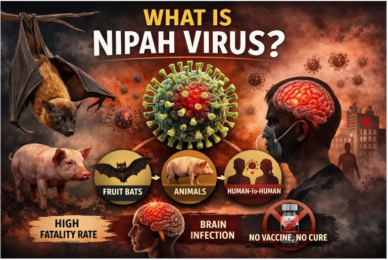
It has not been too long since the world has somewhat recovered from the fear of Covid-19, and now the Nipah Virus has begun to create a new buzz! With a high fatality rate of somewhere between 40 to 75%, Nipah is equally fatal and highly transmissible.
As there is no exact treatment, it’s high time for everyone in the world to know about Nipah, at least its mode of transmission and preliminary signs- as this knowledge can be totally lifesaving! Let’s get into every information we have about the Nipah virus till now and let’s also discover treatment methods that are available for now.
Nipah Virus: Introduction
The natural reservoir of the Nipah virus is Pteropus bats (flying foxes) and it was first known to the world in 1998. During that time, the outbreak was among pig farmers and almost 300 got infected of whom 100 patients died.
Structure: Nipah virus has a genetic material negative sense, single-stranded RNA. It is encapsidated and enveloped and belongs to the Paramyxoviridae family under the Henipavirus genus. The important structure of the Nipah virus everyone should know about is the surface glycoproteins (G and F) as this structure is what helps to adhere to host cells.
How is Nipah Virus Transmitted?
As Nipah virus is a zoonotic disease, its primary source of transmission is bats. When healthy humans get exposed to the fluids and secretions like saliva, urine or even faeces of bats, infection is observed.
Another mode of transmission is an intermediate animal host- commonly pigs. If humans come in close contact with the infected body fluids of animal hosts, successful transmission occurs.
The third and final mode of transmission is human-to-human spread. If a healthy human comes in close contact with the infected person- especially via respiratory droplets, body fluids, or contaminated surfaces (similar to Covid-19), infection occurs. This transmission is what brings about an epidemic or a pandemic.
Signs and Symptoms:
Preliminary Signs
Once the virus enters the body, after a 4 to 14 days of incubation period, initial signs like sudden onset of fever, headache, respiratory illness, vomiting, muscle pain, and breathing difficulty are observed.
Complications
Acute encephalitis: Brain swelling occurs if the patient doesn’t receive timely care or if the immune system is too compromised. This is what makes the disease highly fatal. Once the person enters this phase, he may have to bear uncomfortable signs like persistent headaches, seizures, constant dizziness, altered consciousness, confusion and memory loss.
Depending on the severity of infection, the person may enter a coma within 24 to 48 hours of symptom onset.
Diagnosis
Kits: Simply ELISA or kits utilizing this technique is used for Nipah virus diagnosis. This method has a sensitivity of around 75%. However, this method is only for preliminary identification and for confirmation, molecular techniques like RT-qPCR are recommended.
RT-qPCR: It is a standard test to detect even low copies of viral RNA. This test is more preferred because of its high sensitivity. The test also provides estimates of viral load.
Treatment
- There is no specific treatment for Nipah virus. Only supportive care is available to manage symptoms and prevent complications.
-The recovery is based on the immune status of the infected individual.
- In most people, the mild illness generally resolves within two to four weeks. In cases, the immunity is weak or compromised and cannot fight the virus, the severe complication starts to appear within a short period of time.93 to 21 days after the appearance of initial symptoms).
- The important practices for the infected patients are: consuming nutritious foods and adequate amounts of water, taking rest in isolation, and consuming supporting medications like antipyretics and analgesics.
Prevention and control
-Limit contact with suspected infected individuals. For all pig farmers, they should be highly alert while coming into direct contact with the animals’ body fluids and secretions.
-Hand hygiene is of utmost importance and almost all we can do to prevent infection transmission.
-In cases, Nipah virus goes endemic in your area, early detection and an early health care approach is must to limit the occurrence of encephalitis.
Extras(especially for students):
Pathogenesis
The pathogenesis of Nipah virus is characterized by complex host-virus interaction. Humans and certain animals like pigs and bats are susceptible to Nipah virus infections- in all of these hosts, the basic mode of pathogenesis is the same. All known genotypes of Nipah virus are highly pathogenic, showing variation in virulence and clinical severity at the later stages of the infection.
1. Attachment: The site of binding is the ephrin-B2 and ephrin-B3 receptors which are present and expressed in endothelial, epithelial and smooth muscle cells as well as neurons. At first, the virus attaches and binds to these susceptible cells. The ability of the virus to bind cells of various organs is what explains its ability to infect multiple organs.
2. Internalization: After successfully attaching to the host membrane, the envelope of the virus then fuses with the host cell membrane followed by successful internalization of the viral nucleocapsid.
3. Uncoating and replication: The nuclear material, which is negative sense single stranded RNA cannot bind to the host ribosome for translation. So, the virus starts replication with its own RNA-dependent RNA polymerase.
At first, the negative sense RNA is transcribed into positive sense RNA followed by translation into viral proteins. The replication completes in cytoplasm, and the end result is multiple new viral nucleocapsids.
4. Assembly: The nucleocapsids formed in cytoplasm now assemble with viral proteins present in the cytoplasm- the structure thus formed is a new immature virion that lacks the lipid envelope and surface glycoprotein spikes.
5. Encapsidation: As the virus exits from the infected cell by budding, the nucleocapsid becomes encapsidated, the host-derived lipid envelope is utilized for the process. The viral glycoproteins (G and F) are already inserted in the envelope, synthesized in the endoplasmic reticulum and processed through the Golgi apparatus. Without these glycoproteins, no viral infectivity and attachment can be performed.
The multiplied virus now finds new cells to infect and the whole process is repeated, forming multiple viruses.
References
Singh, R. K., Dhama, K., Chakraborty, S., Tiwari, R., Natesan, S., Khandia, R., Munjal, A., Vora, K. S., Latheef, S. K., Karthik, K., Singh Malik, Y., Singh, R., Chaicumpa, W., & Mourya, D. T. (2019). Nipah virus: epidemiology, pathology, immunobiology and advances in diagnosis, vaccine designing and control strategies - a comprehensive review. The veterinary quarterly, 39(1), 26–55. https://doi.org/10.1080/01652176.2019.1580827
Aditi, & Shariff, M. (2019). Nipah virus infection: A review. Epidemiology and infection, 147, e95. https://doi.org/10.1017/S0950268819000086
https://www.who.int/emergencies/disease-outbreak-news/item/2026-DON593